- Background
- SCR-FL USA Program
- Rotator Cuff and Superior Capsule basics
- Summary
- Upper thigh Fascia Lata autograft
- Graft Preparation
- Post-surgical
- Rehabilitation
- Post-surgical Motion and function recovery
- References
- Patient Stories-SCR's
Superior Capsule Reconstruction or SCR as it is commonly called is an arthroscopic minimally invasive reconstructive procedure for Rotator Cuff tears that are either not repairable or if repairable have a high risk of not healing or retearing. These tears are often described as Massive or Irreparable. Essentially, a large gap has developed and tissue is missing.
Background:
Dr. Teruhisa Mihata MD, PhD in Osaka Japan pioneered the procedure in 2006. Initially he was seeking a solution for his patients who lost the ability to raise their arm because of the Massive/Irreparable tear. Surgeons call this loss pseudo-paralysis of the deltoid, meaning it looks like the deltoid is not working. But in reality, it is that the rotator cuff is no longer providing its key functions and the patient cannot raise the arm without those key functions. Dr Mihata found that he could replace the missing tissue by using a graft of fascia (Fascia Lata) from the upper thigh. This fascia lines muscle and is superficial. It has excellent strength, stiffness, and healing characteristics. It does require a separate superficial incision on the upper thigh to obtain the graft and it does take several months for the graft harvest site to heal and allow return to sport activities. But the results for Dr Mihata have been excellent for the last18 years. He has completed over 1000 and it is a very common procedure in Japan performed by many surgeons with published 10-year success rates of 91%.
The SCR with Fascia Lata (autograft) using Mihata technique is new to the USA as of 2023 even though it was first published in 2013. It is new because the original attempt at SCR in the USA did not use the Fascial Lata Autograft as Dr Mihata recommended. Instead, a dermal allograft with less strength, stiffness and inferior healing characteristics became popular. While it is not entirely clear why surgeons did not follow the Mihata technique, it is very clear that the results using this altered technique were poor. Failure rates of up to 70% have been published.
Dr. Paul B Roache MD, FAANA, FAAOS meet Dr Mihata at a rotator cuff meeting in Zurich in 2019. Dr Roache had research interest in repairing the superior capsule as an important component of a standard rotator cuff tear. Recognizing that Dr Mihata had proven that the superior capsule function alone without a repairable tendon restored rotator cuff function. This mutual interest in the superior capsule led to Dr. Roache visiting Dr Mihata in late 2022 to study the Mihata technique for SCR. Especially, the all-important graft harvest and preparation from the patient’s upper thigh.
In 2023 Dr Roache launched the initial pilot program performing SCR with Fascia Lata autograft. This program had strong supervision and advice from Dr Mihata. In early 2024 the first year was completed with 20 patients undergoing the Mihata technique SCR. The First-year report was reviewed with Dr Mihata when he visited SF for the AAOS meeting. He validated the program and the results. Per his instructions the program is now expanding and will be educating US surgeons to bring the technique to our patients who very much need it here in the USA.


SCR-FL USA Program
The Program has been named: SCR-FL USA also known as Mihata Technique SCR.
Key members of the Program:
Director: Paul B Roache MD, FAAOS, FAANA
Graft surgeon: Arati Dunbar, MD, FAAOS, FAANA *
MRI radiologist: David W. Stoller, MD, FACR
Physical Therapy: Noman Naqvi, PT, DPT
Advisors: Teruhisa Mihata MD, PhD
Akihiko Hasegawa, MD, PhD
* Dr Dunbar is the first US surgeon being trained by Dr Roache in Mihata Technique.
Rotator Cuff and Superior Capsule basics: essential understanding for the Mihata technique.
- The Rotator Cuff is group of 4 muscles and tendons that surround the upper shoulder humeral head and create the shoulder socket.
- The superior capsule is a strong ligamentous structure that supports and compliments the upper 2 tendons of the rotator cuff. (supraspinatus and infraspinatus) it is the equivalent of the subfloor. The tendon is the equivalent of the floor on top of the subfloor.
- The attachment of the Rotator Cuff Tendon and superior capsule to the humerus is 60% superior capsule. Although the superior capsule is thought to compliment the function of the rotator cuff, it may actually be that the rotator cuff compliments the function of the superior capsule. (One reason the Mihata technique is successful, because it restores the 60% attachment of the original anatomy)
- The superior capsule attachment at the humerus connects the anterior tendon of the rotator cuff (the Subscapularis) to the posterior rotator cuff (infraspinatus and teres minor) it does so through a specific structure running from front to back at the lateral insertion to the humerus. This structure in the superior capsule is called the "Rotator Cable" because early surgeons thought it reminded them of the main cable of the Golden Gate Bridge. This Anterior Posterior connection is recreated with the Mihata technique.
- Massive tears are irreparable because they are missing tendon and capsule and because of the gap from the missing tissue are not able to be mobilized for reattachment directly to the bone of the humerus. The graft of the Mihata SCR bridges this gap and provides the subfloor for the remaining tendon and capsule elements to be attached. By attaching to the Graft there is the potential for partial restoration of the function of the muscle and tendon.
- The rotator cuff superior capsule complex has a thickness that fills the space under the acromion. This filling of the space is one of the components lost in massive tears which can alter the ability to raise the arm. A thick graft of the fascia Lata auto graft has been demonstrated to restore this spacing and contribute to the ability to raise the arm.
Summary:
The easiest way to understand the problem of a massive irreparable rotator cuff tendon and superior capsule tear is to think of the rotator cuff as a 3 sided box. The front, the top and the back are connected together through the upper corners of the box. In a massive tear the top and corners have been lost. The SCR with Fascia Lata reconstructs the top of the box and the corners. (The dark box line represents superior capsule)

Upper thigh Fascia Lata autograft:
a 3cm by 12.5 cm strip of fascia is removed. (11/4inch wide by 5 inch long) This requires just under a 15cm (6 inch) incision in the skin to get to the fascia layer which is just under the skin.


Graft preparation:
The strip of fascia is then folded and sutured to create a thick and stiff graft. (to reconstruct the top of the box.)

Post-surgical:
Most patients are eligible to go home after surgery either later in the day or the next morning. This depends on the discomfort for harvesting the graft from the upper thigh. Even though it is very superficial the discomfort can limit immediate ambulation and we Require that the physical therapists confirms you are walking well and doing stairs safely. Most patients have little to no shoulder pain. The anesthesiologist gives a regional anesthetic for the shoulder which keeps discomfort low or away.
Rehabilitation:
Noman Naqvi, PT, DPT is the director of SCR-FL USA rehabilitation, and he is training therapist in other regions of California so patients can travel to a regional SCR-FL USA rehab approved center. Currently, there are PTs in San Francisco County, Napa county, and Mendocino county with planning for San Mateo county underway.
Depending on the specifics of your SCR-FL reconstruction you will be in a sling minimum 4 weeks and up to 8 weeks. Then 3 phases of PT begin.
At 3 months an MRI is obtained and reviewed by our radiologist David W. Stoller, MD FACR with your surgeon Dr. Roache. We are looking for graft thickness and healing to confirm that the more vigorous stages of PT are appropriate to start. The MRI is compared to your Pre-surgical MRI.
- pre-surgery massive tear:
note no space and torn tendon capsule.

- 3-month post SCR-FL MRI: note dark graft tissue and restoration of space

Post-surgical motion and function recovery.
Typical in SCR-FL using Mihata technique.
- 5 month postop Rt sh ROM degre

- ROM 5 months (Rt shoulder)

- 5 month ROM

- 3-month ROM

Patient Stories-SCR's
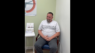
 SCR-FL 8 months postop (patient # 5)
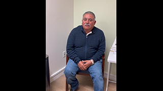
- SCR FL 9 months (patient #4 in Spanish)
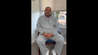
- SCR-FL 11 months (patient #1)
References:
- Clinical results of arthroscopic superior capsule reconstruction for irreparable rotator cuff tears
- Biomechanical Effect of Thickness and Tension of Fascia Lata Graft on Glenohumeral Stability for Superior Capsule Reconstruction in Irreparable Supraspinatus Tears
- Postoperative graft integrity affects clinical outcomes after superior capsule reconstruction using fascia lata autograft in posterior-superior rotator cuff tears: a multicenter study
- Superior Capsule Reconstruction Using Fascia Lata Allograft Compared With Double- and Single-Layer Dermal Allograft: A Biomechanical Study
Other Shoulder Procedures
Rotator Cuff Procedures
- Shoulder Arthroscopy
- Rotator Cuff Repair
- Superior Capsule & Anterior Rotator Cable repair: Recouplage
- REGENETEN Bioinductive Implant
- Rotium Implant
- Proximal Biceps Tenodesis
- Transosseus Arthroscopic Rotator Cuff Repair
- Subacromial Decompression
- Mumford Distal Clavicle Excision
- Revision Rotator Cuff Surgery
- Superior Capsule Reconstruction with Fascia Lata autograft
- In Space Balloon for Irreparable Rotator Cuff Tears
- Latissimus Dorsi Tendon Transfer
- Lower Trapezius Tendon Transfer
Instability Procedures
- Arthroscopic Bankart Repair
- SLAP Repair
- Latarjet Procedure
- Revision Open Labral Repair (Revision Bankart)
- AC Joint Repair
Frozen Shoulder Adhesive Capsulitis Procedures
Shoulder Arthritis Procedures
- Viscosupplementation for Shoulder Arthritis
- Shoulder Joint Replacement
- Reverse Total Shoulder Replacement
- Shoulder Resurfacing
Fracture Shoulder Procedures
- ORIF of the Clavicle Fractures
- Proximal Humerus Fractures
- ORIF of the Scapula Fractures
- Complex Fracture Repair of the Shoulder
- Nonunion Fixation of a Shoulder Fracture
- Malunion Surgery of the Shoulder
- Periprosthetic Shoulder Fracture Fixation